
FIGURE
Fig. 7
- ID
- ZDB-FIG-190305-7
- Publication
- LaBonty et al., 2018 - Injury of Adult Zebrafish Expressing Acvr1lQ204D Does Not Result in Heterotopic Ossification.
- Other Figures
- All Figure Page
- Back to All Figure Page
Fig. 7
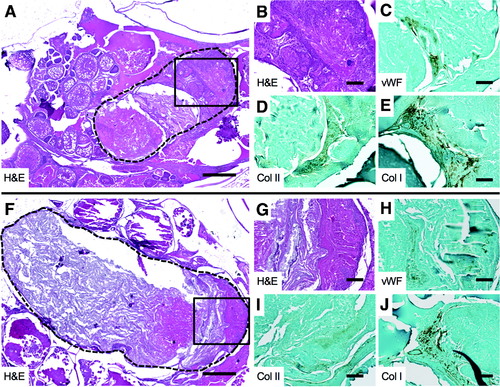
Cartilage and bone formation in body cavity HO in Acvr1lQ204D-expressing zebrafish. HO lesions in tail fin clip-injured HS Tg(Bre:GFP); Tg(acvr1l_Q204D-mCherry) zebrafish at 2 wpi, paraffin sectioned and H&E stained (A, B, F, G), and analyzed by IHC for vWF (C, H), Col II (D, I), and Col I (E, J). The HO lesion is outlined with a dashed line(A, F). (B, G) Enlarged views of boxed areas in (A, F), respectively. (A, F) 5 × scale bar is 40 μm. (B–E, G–J) 20 × scale bar is 100 μm. Col I, collagen I; Col II, collagen II; vWF, von Willebrand factor. |
Expression Data
Expression Detail
Antibody Labeling
Phenotype Data
| Fish: | |
|---|---|
| Condition: | |
| Observed In: | |
| Stage: | Adult |
Phenotype Detail
Acknowledgments
This image is the copyrighted work of the attributed author or publisher, and
ZFIN has permission only to display this image to its users.
Additional permissions should be obtained from the applicable author or publisher of the image.
Full text @ Zebrafish