
Fig. 5
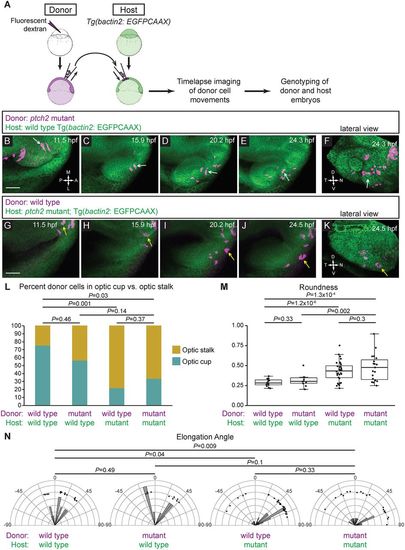
ptch2tc294z acts in a non-cell-autonomous manner to disrupt cell movements. (A) Schematic of cell-transplantation approach. Cells were transplanted from donor to host at blastula stages, and time-lapse microscopy performed on embryos in which transplanted cells were in the prospective optic fissure forming region at 12 hpf. After imaging, both donor and host embryos were genotyped. (B-F) Transplantation of ptch2tc294z mutant cells into a wild-type host. (B-E) Mutant cells exhibit an elongated morphology, move out of the midline region, through the optic stalk, and into the optic cup, where a mutant donor cell contributes to the optic fissure (white arrows). (F) Lateral view of 3D rendering, final time point. White arrow marks a cell that contributes directly to the optic fissure margin. (G-K) Transplantation of wild-type cells into a ptch2tc294z mutant host. (G-J) Wild-type cells move out of the midline region; few cells move to the central retina within the optic cup. Most cells become less elongated and reside in the optic stalk region (yellow arrows). (K) Lateral view of 3D rendering, final time point. Yellow arrow marks cells that arose from the prospective optic cup/fissure region, but contribute to the optic stalk. (L-N) Quantification of transplantation results. n=14 cells (wt into wt); 10 cells (mut into wt); 32 cells (wt into mut); and 19 cells (mut into mut). (L) Proportions of transplanted cells contributing to optic stalk (gold) or optic cup/fissure (teal). Fisher's exact test. (M) Roundness of transplanted cells: morphology reflects host genotype. Unpaired Welch's t-test to account for unequal variance. (N) Elongation angle of transplanted cells. F-test to determine the probability that the variances are not significantly different. Scale bars: 50 µm. |