
Fig. 2
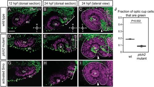
Extended evagination cell movements are disrupted by overactive Hh signaling. (A-I) Wild-type (A-C), ptch2tc294z mutant (D-F) and activated Smo RNA-injected (G-I) embryos subjected to nls-Kaede photoconversion at the optic vesicle stage (A,D,G). The entire optic vesicle was converted from green to red (magenta) fluorescence (along with some ectoderm). (B,E,H) Single confocal sections of optic cups from photoconverted embryos, 24 hpf, dorsal view. (C,F,I) 3D-rendered optic cups, 24 hpf, lateral view. Dashed lines outline the optic cup. Arrowheads indicate the optic fissure. (J) Quantification of extended evagination movements, indicated by the proportion of green nuclei in the optic cup (green nuclei divided by the total number of nuclei) in wild-type (wt) and ptch2tc294z mutant embryos. The proportion of green nuclei in the optic cup is significantly reduced in ptch2tc294z mutants. n=3 wt, 2 mut. Unpaired Student's t-test. ec, ectoderm; le, lens; os, optic stalk; ov, optic vesicle. Scale bar: 50 µm. |
| Fish: | |
|---|---|
| Observed In: | |
| Stage: | Prim-5 |