
Fig. 4
- ID
- ZDB-FIG-190129-9
- Publication
- Goldshmit et al., 2018 - Different Fgfs have distinct roles in regulating neurogenesis after spinal cord injury in zebrafish
- Other Figures
- All Figure Page
- Back to All Figure Page
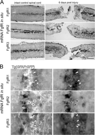
FgfRs expression around the central canal of the spinal cord on GFAP expressing radial glia. a Longitudinal sections show FgfR1–3 mRNA expression in cells at the central canal in intact uninjured spinal cord. At 6 days post injury (dpi) in situ hybridization shows an increase of the mRNA of all three FgfRs in these central canal cells particularly around the lesion site. b Sections from spinal cords in Tg(gfap:EGFP) transgenic fish show the location of GFAP+ ependymal radial glia cells and in situ hybridisation mRNA signal for FgfRs 1–3. At least some of the glia cells express varying levels of particularly FgfR1 and FgfR3 (arrowheads) with little overlap observed for FgfR2 (asterisks). Scale bar in A is 200 μm, Scale bar in B is 50 μm |