
Fig. 5
- ID
- ZDB-FIG-190129-10
- Publication
- Goldshmit et al., 2018 - Different Fgfs have distinct roles in regulating neurogenesis after spinal cord injury in zebrafish
- Other Figures
- All Figure Page
- Back to All Figure Page
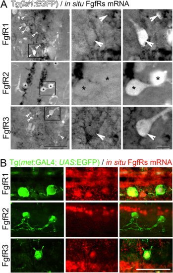
FgfRs expression in cells around the central canal and Islet1 and c-Met expressing motor neurons 6 days post spinal cord injury. a Sections from Tg(Isl1:GFP) transgenic fish showing the location of Islet1+ motor neurons compared to in situ hybridisation for FgfRs 1–3 mRNA. The middle and left panels are higher magnification insets of the boxes indicated in the left panels showing either FgfR signal alone (middle) or merged channels (right). At least some of the Islet1+ motor neurons express FgfR1 and 3 (arrowheads), but not FgfR2 (asterisks). b Sections from Tg(met:GAL4; UAS:EGFP) transgenic fish showing the location of c-Met+ motor neurons compared to in situ hybridisation for FgfRs 1–3 mRNA. Similarly as above, C-met neurons co-labelled with FgfR1 and 3, but not FgfR2 mRNA. The right panels show the merged C-met only (green - left panels) and in situ FgfR mRNA only (red - middle panels). Scale bar in A for left panels is 50 μm, and for middle and right panels is 10 μm. Scale bar in B is 50 μm |