|
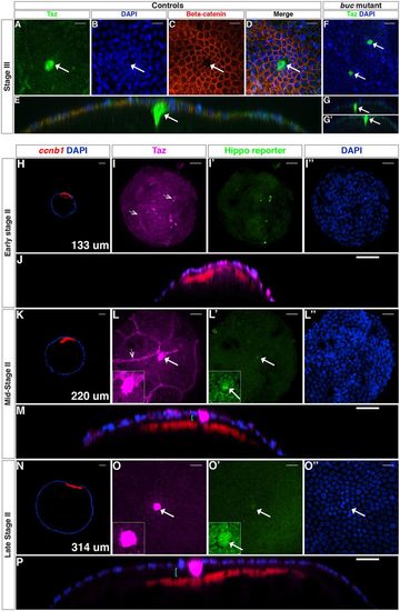
Taz protein is a bona fide marker of the MC. (A-E) Confocal images of stage III WT oocytes stained with anti-Taz (A) and anti-β-catenin (C) antibodies, and DAPI (B, n=4). D shows the three channels merged, and E is an orthogonal view of D. (F-G′) Stage III buc mutant oocytes stained with the anti-Taz antibody and DAPI (n=4). G and G′ are orthogonal views of F. (H-P) Confocal images of wholemount Tg(4xGTIIC:d2egfp)mw50/0 oocytes stained with the anti-Taz antibody (magenta), ccnb1 mRNA (red) and DAPI (blue) at early stage II (H-J), mid-stage II (K-M) and late stage II (N-P) (n=38). Hippo reporter activity is shown in green. Cyan brackets in orthogonal views (M,P) indicate the gap between the oocyte surface and the FC layer (microvilli are not visible); white arrows indicate an MC; dotted arrowheads (I,L) point to non-follicle cells. Insets in L,L′,O,O′ show higher magnification of center of panel. Scale bars: 20 μm in A-D,F,I,J,L,M,O,P; 40 μm in H,K,N.
|