
FIGURE
Fig. S10
- ID
- ZDB-FIG-190114-19
- Publication
- Olcese et al., 2017 - X-linked primary ciliary dyskinesia due to mutations in the cytoplasmic axonemal dynein assembly factor PIH1D3
- Other Figures
- All Figure Page
- Back to All Figure Page
Fig. S10
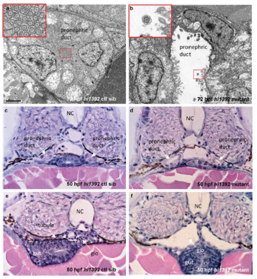
Pronephric cyst phenotypes in zebrafish hi1392Tg mutants (a, b) Transmission electron microscopy at 72 hours post fertilization shows dilation of the pronephros in hi1392Tg mutants (b) and disruption of the usual tightly packed arrangement of motile cilia that is seen in controls siblings (ctl sib, a). Scale bar, 2 μm. Insets are higher magnification images of the indicated pronephric cilia. (c-f) Cross sections of hematoxylin and eosin-stained 50 hpf embryos show the dilated pronephric ducts and glomeruli of mutants (d, f) compared to control siblings (c, e). NC, notochord; glo, glomerulus. |
Expression Data
Expression Detail
Antibody Labeling
Phenotype Data
| Fish: | |
|---|---|
| Observed In: | |
| Stage Range: | Long-pec to Protruding-mouth |
Phenotype Detail
Acknowledgments
This image is the copyrighted work of the attributed author or publisher, and
ZFIN has permission only to display this image to its users.
Additional permissions should be obtained from the applicable author or publisher of the image.
Full text @ Nat. Commun.