
|
Fig. S10
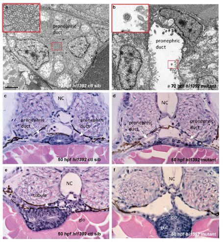
Pronephric cyst phenotypes in zebrafish hi1392Tg mutants
(a, b) Transmission electron microscopy at 72 hours post fertilization shows dilation of the pronephros in hi1392Tg mutants (b) and disruption of the usual tightly packed arrangement of motile cilia that is seen in controls siblings (ctl sib, a). Scale bar, 2 μm. Insets are higher magnification images of the indicated pronephric cilia. (c-f) Cross sections of hematoxylin and eosin-stained 50 hpf embryos show the dilated pronephric ducts and glomeruli of mutants (d, f) compared to control siblings (c, e). NC, notochord; glo, glomerulus.