
Fig. 6
- ID
- ZDB-FIG-190108-39
- Publication
- Krishnakumar et al., 2018 - Functional equivalence of germ plasm organizers
- Other Figures
- All Figure Page
- Back to All Figure Page
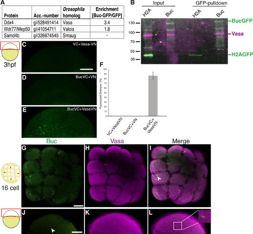
Buc binds zebrafish Vasa. (A) Zebrafish homologs of known Oskar binding proteins in the Buc-interactome detected Vasa (Ddx4) and Valois (Wdr77/Mep50), but not Smaug (Samd4b). Enrichment indicates the ratio of unique peptide counts after Buc-GFP pulldown to GFP-control samples. (B) Buc binds to Vasa in vivo during germ cell specification. Immunoprecipitations from 3 hpf H2A-GFP (42 kD) or Buc-GFP (130 kD) transgenic embryos blotted with GFP (green) and Vasa (magenta) (input = 20% of pulldown). (C-F) Buc and Vasa interact in bimolecular fluorescent complementation assays (BiFC). (C-E) live embryos at 3 hpf as indicated by the cartoon on the left, are not fluorescent (green) upon injection of mRNA encoding VC with Vasa-VN (C; 0±0%; n = 67) or Buc-VC with VN (D; 0±0%; n = 56), but form fluorescent Venus protein with Buc-VC and Vasa-VN (E; 86.5±7.5%; n = 53). Scale bar (C-E): 100 μm. (F) Quantification of BiFC assay (three independent experiments for each RNA). Error bars represent standard deviation of the mean. (G-L) Immunostaining of 16-cell stage (G-I) or 3 hpf (J-L) embryo as indicated by the cartoon on the left showing expression of Buc (green) and Vasa (magenta), inset in (L) shows a 10x magnification of the boxed area. Scale bar (G-L): 200μm. |