
Fig. 2
- ID
- ZDB-FIG-181206-8
- Publication
- Ahlberg et al., 2018 - Rare truncating variants in the sarcomeric protein titin associate with familial and early-onset atrial fibrillation
- Other Figures
- All Figure Page
- Back to All Figure Page
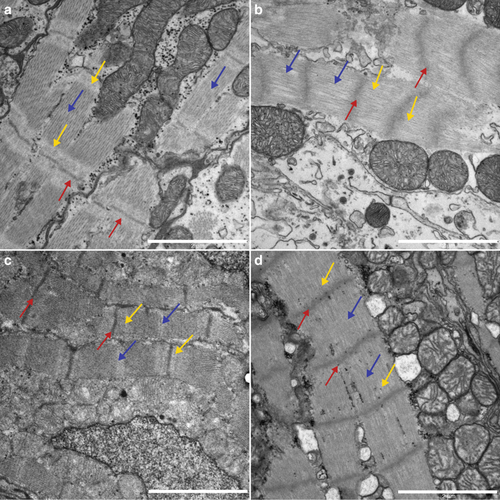
Compromised sarcomere structure in adult heterozygous zebrafish mutants. TEM images (13,500 × ) from adult atria (a, b) and ventricles (c, d). a WT atria show well-defined sarcomeres, with distinguishable Z-discs (red arrows) and I-bands (yellow arrows) throughout the tissue. Scale bar 2 µm. b In heterozygous mutant atria the sarcomere structure is less organized. The Z-discs appear blurred (red arrows), and the I-bands are absent (yellow arrows). Scale bar 2 µm. c Ventricle sarcomeres appear well defined in WT siblings, with clear Z-discs (red arrows), I-bands (yellow arrows), and M-lines (blue arrow). Scale bar; 2 µm. d In heterozygous mutants, there is a distinct lack of I-bands and M-lines, and the Z-discs appear blurry and increased in thickness (Scale bar; 2 µm). The length of the sarcomeres, as measured from Z-disc to Z-disc, are significant different between WT and heterozygous in atria |
| Fish: | |
|---|---|
| Observed In: | |
| Stage: | Adult |