
- Title
-
Rare truncating variants in the sarcomeric protein titin associate with familial and early-onset atrial fibrillation
- Authors
- Ahlberg, G., Refsgaard, L., Lundegaard, P.R., Andreasen, L., Ranthe, M.F., Linscheid, N., Nielsen, J.B., Melbye, M., Haunsø, S., Sajadieh, A., Camp, L., Olesen, S.P., Rasmussen, S., Lundby, A., Ellinor, P.T., Holst, A.G., Svendsen, J.H., Olesen, M.S.
- Source
- Full text @ Nat. Commun.
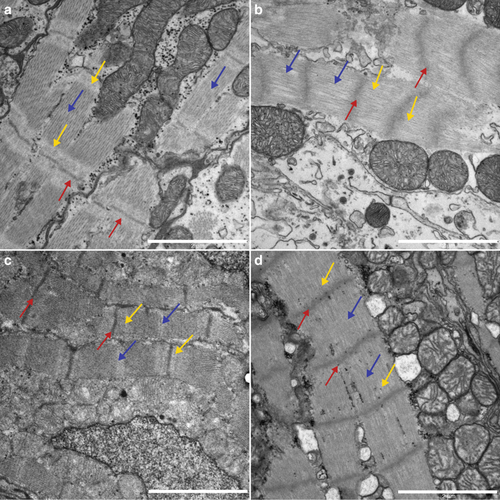
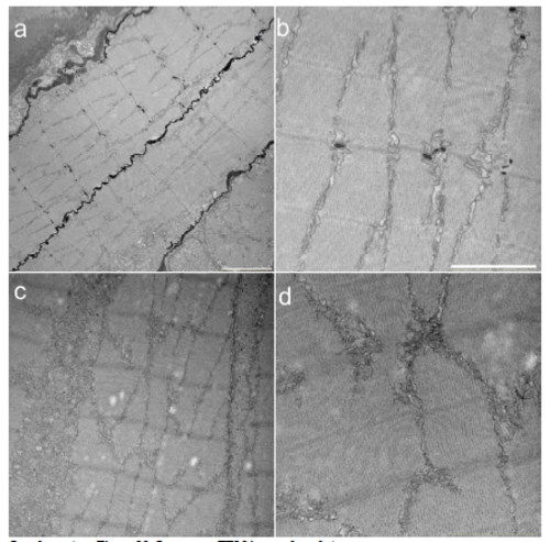
Compromised sarcomere structure in adult heterozygous zebrafish mutants. TEM images (13,500 × ) from adult atria (a, b) and ventricles (c, d). a WT atria show well-defined sarcomeres, with distinguishable Z-discs (red arrows) and I-bands (yellow arrows) throughout the tissue. Scale bar 2 µm. b In heterozygous mutant atria the sarcomere structure is less organized. The Z-discs appear blurred (red arrows), and the I-bands are absent (yellow arrows). Scale bar 2 µm. c Ventricle sarcomeres appear well defined in WT siblings, with clear Z-discs (red arrows), I-bands (yellow arrows), and M-lines (blue arrow). Scale bar; 2 µm. d In heterozygous mutants, there is a distinct lack of I-bands and M-lines, and the Z-discs appear blurry and increased in thickness (Scale bar; 2 µm). The length of the sarcomeres, as measured from Z-disc to Z-disc, are significant different between WT and heterozygous in atria PHENOTYPE:
|
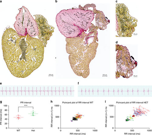
Truncating mutation in ttn.2 cause increase fibrosis and electrophysiological defects in adult heterozygous mutants. Sirius staining of isolated whole hearts from adult WT (a) and heterozygous mutant siblings (b) show increased fibrotic lesions in the heterozygous heart (b) compared with the WT (a). This increase appears to be more pronounced in the atria of the heterozygous hearts (d) (n = 4) compared with that of the WT siblings (c) (n = 4). Scale bars: a, b 200 µm; c, d; 50 µm. ECG surface recordings of adult WT fish (e) revealed a regular ECG pattern with well-defined P-waves and QRS complexes, with regular PR intervals (g) and RR intervals, with low beat-to-beat variability, as shown by the Poincaré plot (h), indicative of a regular heart rhythm (n = 8). In the heterozygous siblings, the ECG pattern was equally well defined (f), but with a larger PR interval, compared with the WT siblings. Furthermore, the RR interval and beat-to-beat variability was irregular, as demonstrated by the Poincaré plot (i), indicating an irregular heart rhythm in the heterozygous siblings (n = 8) PHENOTYPE:
|
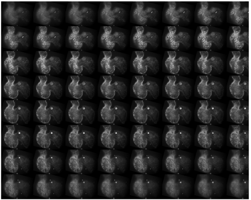
Montage of Z-stack images, wild type (WT) Zebrafish Complete Z-stack images of the WT zebrafish hearts shown in the manuscript’s figure 3 |
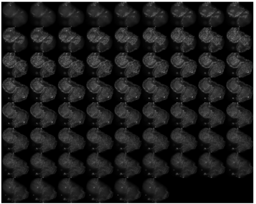
Montage of Z-stack images, mutant zebrafish Complete Z-stack images of the mutant zebrafish hearts shown in the manuscript’s figure 3 |
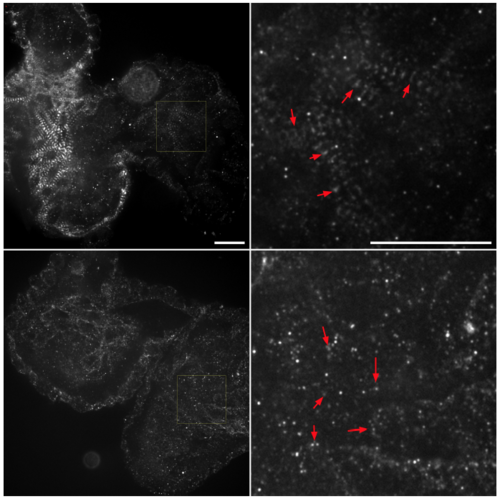
Defective z-discs in a zebrafish titin truncated homozygous mutant Top panel) Isolated hearts form 72 hours post fertilization old zebrafish stained for alpha-actinin, an important component of the Z-disc, revealed well-defined z-discs in both the atria (right chamber) and ventricle (left chamber) in the WT siblings. A close up of the atria (right) show well-defined z-discs (red arrows). Bottom panel) Compared to the observations made in the isolated hearts from the WT sibling embryos, the homozogous mutant embryos showed a no well-defined z-discs, and an aberrant alpha-actinin expression in the atria (indicated by the red arrows in the bottom right image) and the ventricle indicating a compromised sarcomere architecture. Scale bars = 20μm. Please see supplementary Figure 9-10 for complete z-stacks. EXPRESSION / LABELING:
PHENOTYPE:
|
Sarcomere TEM images larval stage TEM images from 3 dpf ttn.2sfc9+/+ (a-b) and ttn2.sfc9+/- (c-d) larvae hearts. a) show well-defined sarcomeres, with distinguishable Z-discs with adjacent I-bands, and distinct M-lines throughout the cardiac tissue. Scale bar 2μM (a)and 4 μM (b). In heterozygous mutant larvae (c) the sarcomere structure is less organised. The z-discs appear blurred and the I-bands are absent (d) Scale bar 2μM (c) and 4 μM respectively(d) PHENOTYPE:
|

ZFIN is incorporating published figure images and captions as part of an ongoing project. Figures from some publications have not yet been curated, or are not available for display because of copyright restrictions. PHENOTYPE:
|
|
ZFIN is incorporating published figure images and captions as part of an ongoing project. Figures from some publications have not yet been curated, or are not available for display because of copyright restrictions. PHENOTYPE:
|