
Fig. S4
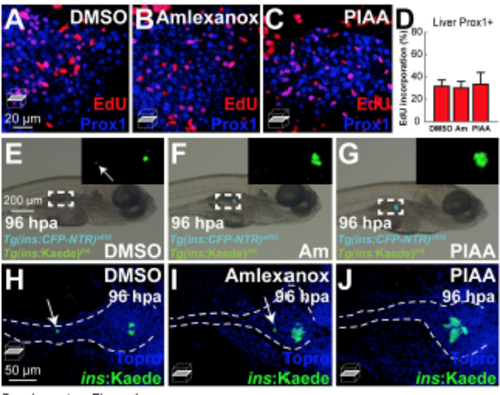
TBK1/IKKε inhibitors do not increase proliferation of liver cells nor lead to an overshoot in b-cell number. (A-C) Confocal single-plane images of [Tg(ins:CFP-NTR)s892; Tg(ins:Kaede)jh6] larvae at 48 hpa, concurrently treated with EdU and DMSO (A), amlexanox (B), or PIAA (C), respectively, from 0-48 hpa, stained for Prox1 (blue). The number of Prox1- positive cells in the liver that incorporated EdU did not significantly increase in TBK1/IKKε-Itreated recovering larvae (B and C) compared to DMSO-treated larvae (A). (D) The percentage (mean±SD) of Prox1-positive liver cells that incorporated EdU at 48 hpa (in A-C; 31.6±5.6% (DMSO), 30.1±5.8% (amlexanox), and 33.5±10.5% (PIAA)). Cells in 10 planes of confocal images from 5 individual larvae were counted per condition. (E-G) Bright-field images combined with fluorescent images showing the overall morphology of embryos and [Tg(ins:CFP-NTR)s892; Tg(ins:Kaede)jh6] expression (green) in larvae at 96 hpa treated with DMSO (E), amlexanox (F), and PIAA (G), respectively. While TBK1/IKKε-Is expanded [Tg(ins:CFP-NTR)s892; Tg(ins:Kaede)jh6]-expressing cell population (white squares and insets) during regeneration (F and G) compared to DMSO (E), a longer treatment (0-96 hpa) did not result in overproliferation of b-cells. (H-J) Confocal single-plane images of [Tg(ins:CFPNTR) s892; Tg(ins:Kaede)jh6] larvae at 96 hpa (in E-G), stained with Topro (blue). White arrows indicate b-cells located in secondary islets. |