
Fig. 6
- ID
- ZDB-FIG-181108-16
- Publication
- Ranski et al., 2018 - Characterization of retinal regeneration in adult zebrafish following multiple rounds of phototoxic lesion
- Other Figures
- All Figure Page
- Back to All Figure Page
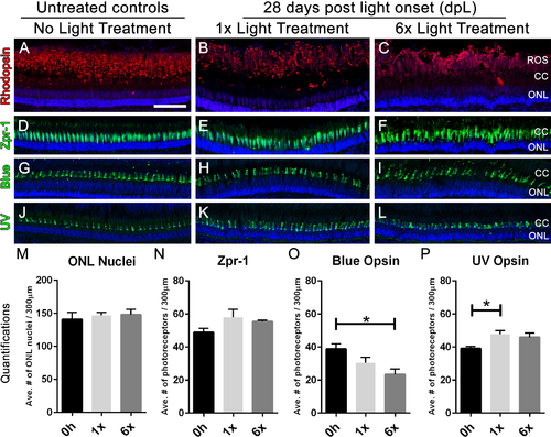
Following six rounds of light treatment, retinas replace the lost photoreceptors.(A–L) Retinal sections collected at 28 dpL immunolabeled with anti-rhodopsin, Zpr-1, anti-blue opsin, and anti-UV opsin to show regeneration of the photoreceptors in untreated control (No Light Treatment), experimental control (1×Light Treatment), and experimental retinas (1×Light Treatment) following photolytic damage. S Nuclei are stained blue with TO-PRO-3. (A–C). Rod photoreceptor outer segments are immunolabeled with anti-rhodopsin (red). (D–F) Red-green double cones are immunolabeled with Zpr-1 (green). (G–I) Long single cones are immunolabeled with anti-blue opsin (green). (J–L) Short single cones are immunolabeled with anti-UV opsin (green). (M–P) Quantification of the average number of photoreceptors 28 dpL in control and experimental groups (n = 5). Cells were counted over a linear distance of 300 µm on the central dorsal retina. Single asterisks indicate significant differences between groups (p < 0.03). Scale bar represents 25 μm. |
| Fish: | |
|---|---|
| Condition: | |
| Observed In: | |
| Stage: | Adult |