
Fig. S3
- ID
- ZDB-FIG-181024-3
- Publication
- Grassini et al., 2018 - Nppa and Nppb act redundantly during zebrafish cardiac development to confine AVC marker expression and reduce cardiac jelly volume
- Other Figures
- All Figure Page
- Back to All Figure Page
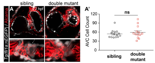
No increase in endocardial cell number is observed in nppa/nppb double mutants compared with siblings. A. Confocal stacks of 72 hpf sibling and double mutant with endocardial nuclei labelled by Tg(fli1a:nEGFP) (white) and myocardial membranes labelled using an anti-Alcama antibody (red). In addition to myocardium, endocardial cushions are also labeled with Alcama, distinguishing them from the remaining endocardium. Double positive cells (white arrows) are quantified in A’. A’. Dot plots representing the number of AVC cells in sibling (n = 14) versus double mutants (n = 13), showing no significant difference (ns; twotailed t-test; error bars indicate the standard error mean). |