
Fig. S1
- ID
- ZDB-FIG-181018-18
- Publication
- Du et al., 2018 - A transgenic zebrafish model for in vivo long-term imaging of retinotectal synaptogenesis
- Other Figures
- All Figure Page
- Back to All Figure Page
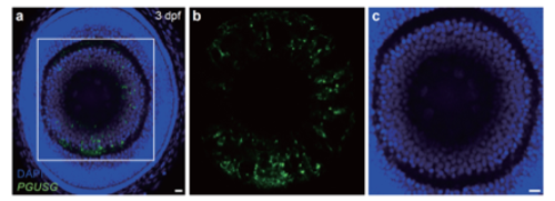
DAPI staining of the retina in PGUSG larvae. (a) One optical section of confocal image of the retina with DAPI staining in a 3-dpf PGUSG larva. The nasal is upwards, the dorsal is leftwards. (b,c) Zoom-in and color separate view of the boxed region in (a). We chose three sections of each retina to count the number of Sypb-EGFP positive RGCs and DAPI-stained RGCs. Taking DAPI-stained RGCs as total RGCs, we found that 14.5 ± 0.5% (mean ± s.e.m.) of the RGCs were EGFP-positive in PGUSG at 3 dpf. As ~ 50% of RGCs could be labelled by the pou4f3 driver25, our PGUSG lines can at most label ~ 30% of pou4f3-positive RGCs. The calculation was based on data obtained from 3 larvae. Scale bar, 5 μm. |