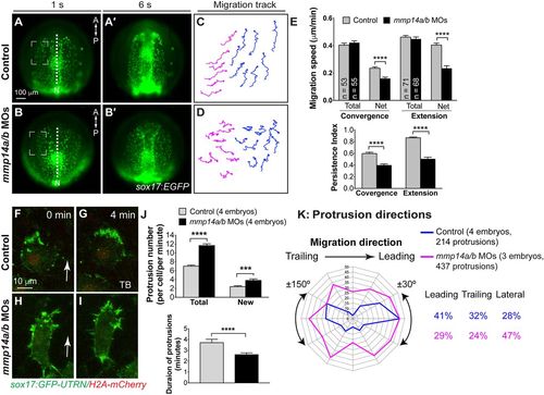
Mmp14a and Mmp14b are required for convergence and extension movements of the anterior endodermal cells. (A-E) Epifluorescence time-lapse experiments for indicated embryos (Movie 6). (A-B′) Still images from movies at 1s and 6s. Dashed squares denote regions in which cells were analyzed. A, anterior; P, posterior. (C,D) Representative migration tracks of anterior endodermal cells in A and B. Blue and magenta tracks represent cells that migrated primarily in the anterior and medial directions, respectively. (E) Total and net speeds of convergence and extension movements, persistence index of cell migration, for the entire lengths of movies, in the embryos indicated (five embryos per group). The numbers of cells analyzed are indicated in the graph. ****P<0.0001; Student's t-test. (F-K) Actin dynamics as assessed by confocal time-lapse imaging of anterior endodermal cells expressing GFP-UTRN in the embryos indicated. (F-I) Representative confocal still images at 0 and 4 min (Movie 7). Arrows indicate direction of migration. (J) Total number (in each endodermal cell at 1 min intervals for the imaging period, 671 protrusions from six control cells, 926 protrusions from six morphant cells), newly formed protrusions (in each endodermal cell per minute, 198 protrusions in six control cells and 280 protrusions in six morphant cells) and duration of protrusions (35 protrusions from six control cells, 34 protrusions from six morphant cells). ***P<0.001; ****P<0.0001; Student's t-test. (K) Direction of protrusions relative to the direction of cell migration in sibling and mmp14a/b MOs-injected embryos (2 min intervals, grouped into 30° sectors). Percentage of protrusions in different directions (leading, ±30°; trailing, ±150°; lateral, ±30-150°) is shown.
|
