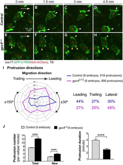
Glypican 4 is required to maintain polarized actin-rich protrusions on migrating endodermal cells. Actin dynamics were assessed by tracking endodermal cells expressing GFP-UTRN (Movie 2). (A-H) Snapshots from confocal time-lapse imaging at different time points. Broader lamellipodia are marked by white arrowheads (control cells) and smaller lamellipodia by yellow arrowheads (gpc4 mutant cells). White arrows indicate the direction of migration of endodermal cells. (I) Direction of protrusions relative to the direction of cell migration in sibling and gpc4 mutant embryos (2 min intervals, grouped into 30° sectors). Percentage of protrusions in various directions (leading, ±30°; trailing, ±150°; lateral, ±30-150°) is shown. (J) Average total protrusions (in each endodermal cell, as assessed at 1 min intervals throughout the imaging period, 1286 protrusions in 11 control cells, 1091 protrusions in eight mutant cells), newly formed protrusions (in each endodermal cell per minute, 443 protrusions in 11 control cells, 326 protrusions in six mutant cells) and the duration of protrusions in control (50 protrusions, 11 cells) and gpc4 mutants (31 protrusions, eight cells). ****P<0.0001; Student's t-test.
|
