
FIGURE
Fig. 2
Fig. 2
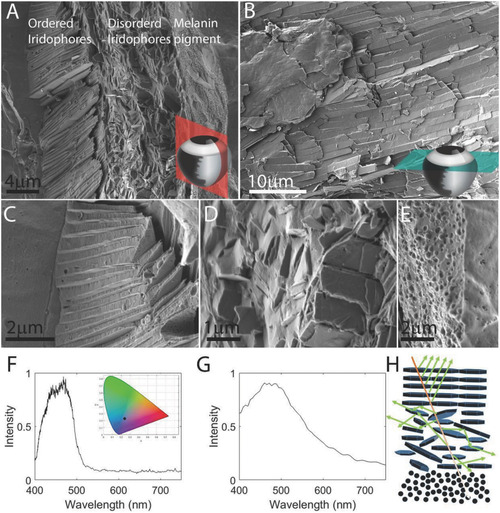
The ultrastructure and optical properties of the iris. A) A coronal section through the iris showing three distinct layers: (i) ordered iridophores, (ii) disordered iridophores, and (iii) a pigmented layer. The section was taken from the periphery of the eye where the iris is curved and, thus, the crystals in the ordered layer are tilted. B) A top view of the iris obtained by a transverse section through the eye shows the elongated guanine crystals packed one next to the other, perfectly tiling the surface of the iris. The anatomical location of sections is illustrated in the bottom‐right corners in (A) and (B). C–E) Higher magnifications of the different iris layers: ordered iridophores, disordered iridophores, and pigmented layer. A–E) Cryo‐SEM images of high‐pressure frozen, freeze‐fractured iris. F) Graph showing measured reflectance from the iris, revealing a broad peak centered around 450 nm. Inset shows the color of reflected light (black dot) plotted on a 1931 CIE chromaticity space diagram. G) Simulated reflectance of the ordered layer calculated based on the data in high‐resolution cryo‐SEM images. H) A sketch illustrating the different paths of light traveling through the multilayered iris. Scale bars: 4 µm (A), 10 µm (B), 2 µm (C,E), 1 µm (D).
|
Expression Data
Expression Detail
Antibody Labeling
Phenotype Data
Phenotype Detail
Acknowledgments
This image is the copyrighted work of the attributed author or publisher, and
ZFIN has permission only to display this image to its users.
Additional permissions should be obtained from the applicable author or publisher of the image.
Full text @ Adv Sci (Weinh)