
FIGURE
Fig. S3
Fig. S3
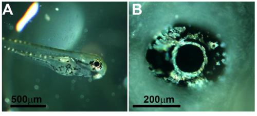
Light microscopy images of 3 dpf zebrafish larvae. (A) A complete larva. (B) Magnification of the eye. Already at this stage, the iridophores in the larval iris are clearly visible. The iridophores surrounding the lens seem to be the first to form. Scale bars: 500 μm (A), 200 μm (B). |
Expression Data
Expression Detail
Antibody Labeling
Phenotype Data
Phenotype Detail
Acknowledgments
This image is the copyrighted work of the attributed author or publisher, and
ZFIN has permission only to display this image to its users.
Additional permissions should be obtained from the applicable author or publisher of the image.
Full text @ Adv Sci (Weinh)