
FIGURE
Fig. S9
- ID
- ZDB-FIG-180918-32
- Publication
- Xing et al., 2018 - Mutational analysis of dishevelled genes in zebrafish reveals distinct functions in embryonic patterning and gastrulation cell movements
- Other Figures
- All Figure Page
- Back to All Figure Page
Fig. S9
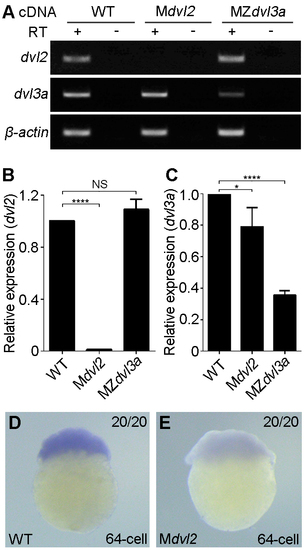
Analysis of mutant dvl2 and dvl3a transcripts. (A) Semi-quantitative RT-PCR analysis to detect mutant dvl2 and dvl3a transcripts at 1-cell stage. ß-actin served as a loading control. NMD can be observed for dvl2 and dvl3a transcripts, respectively. (B, C) Quantification of mutant dvl2 and dvl3a mRNA levels in Mdvl2 and MZdvl3a mutants. The expression level in WT embryo is set as 1 after normalization with ß-actin. Bars represent the mean ± s.d. from three experiments (*, P<0.05; ****, P<0.0001). (D, E) In situ hybridization analysis of dvl2 transcripts in WT and Mdvl2 embryos. RT, reverse transcriptase. |
Expression Data
Expression Detail
Antibody Labeling
Phenotype Data
Phenotype Detail
Acknowledgments
This image is the copyrighted work of the attributed author or publisher, and
ZFIN has permission only to display this image to its users.
Additional permissions should be obtained from the applicable author or publisher of the image.
Full text @ PLoS Genet.