
Fig. S5
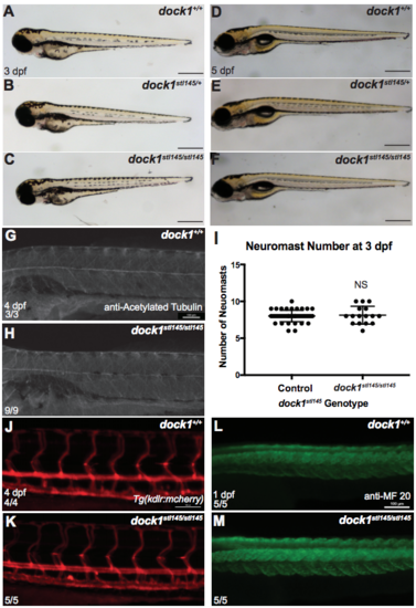
Gross development is normal at 3 dpf comparing A) wild-type, B) heterozygous, and C) mutant larvae from a dock1stl145 intercross. Scale bars = 500 μm. D-F) Gross development is normal and swim bladders have inflated at 5 dpf comparing D) wild-type, E) heterozygous, and F) mutant from a dock1stl145 intercross. Scale bars = 500 μm. G) Acetylated tubulin shows axons are present and well-fasiculated in both wild-type (n = 3) and H) dock1stl145/stl145 mutant larvae (n = 9) at 4 dpf. I) Neuromast number, detected by DASPEI labeling, did not vary between controls (n = 46) or mutants (n = 16) at 3 dpf (NS, p = 0.7518), indicating that global PLLn development is not affected. Bars represent means ± SD; unpaired t Test with Welch’s correction. J) Tg(kdlr:mcherry) labeling blood vessels at 4 dpf in wild-type and K) dock1stl14/stl145 mutants. L) MF 20 staining shows defined somite development in wild-type and M) dock1stl14/stl145 mutant larvae at 1 dpf. Scale bars = 100 μm. (PDF 5492 kb) |