
Fig. S5
- ID
- ZDB-FIG-180912-11
- Publication
- Gurung et al., 2018 - Distinct roles for the cell adhesion molecule Contactin2 in the development and function of neural circuits in zebrafish
- Other Figures
- All Figure Page
- Back to All Figure Page
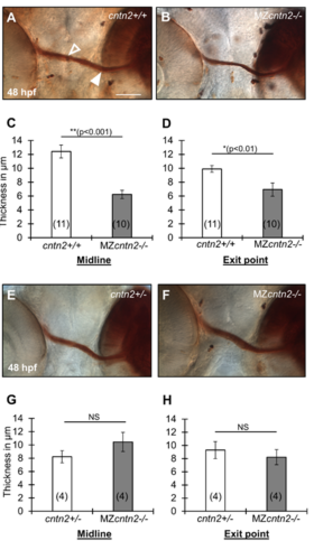
Retinal ganglion cell axon fascicles are variably affected in cntn2 mutants (A, B, E, F) Ventral view of DiI-labeled retinal ganglion cell (RGC) axon fascicles after photoconversion in cntn2+/+ (A), cntn2+/- (E) and MZcntn2-/- (B, F) embryos at 48 hpf. The DiI-injected region targeting the nasal RGCs appears as a dark brown area on the right side in each panel. The filled arrowhead marks an RGC axon fascicle at the exit point from the eye, and the open arrowhead marks an RGC axon fascicle at the midline. (C, D, G, H) Summary of RGC axon fascicles thickness in wild-type and MZcntn2-/- embryos. The RGC axon fascicles are significantly thinner at the midline (C) and at the exit point from the eye (D) in MZcntn2-/- mutants (B) compared to cntn2+/+ cousins (A). However, these differences were not seen (G, H) when MZcntn2-/- mutants (F) were compared to cntn2+/- siblings (E). Scale bar in A, 50 μm for A, B, E, and F. Unpaired t-test; NS: not significant. Error bars show Mean ± SEM. |
Reprinted from Mechanisms of Development, 152, Gurung, S., Asante, E., Hummel, D., Williams, A., Feldman-Schultz, O., Halloran, M.C., Sittaramane, V., Chandrasekhar, A., Distinct roles for the cell adhesion molecule Contactin2 in the development and function of neural circuits in zebrafish, 1-12, Copyright (2018) with permission from Elsevier. Full text @ Mech. Dev.