
FIGURE
Fig. 3
Fig. 3
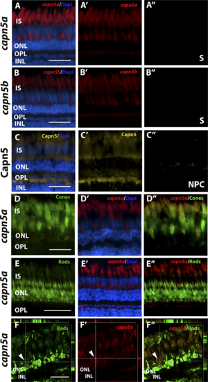
Capn5 is expressed specifically in the cone photoreceptors of the adult zebrafish retina. (A–B″) FISH showing expression of capn5a and capn5b in the adult (WT) retina; expression was seen in the inner segments of the cones. (A″) and (B″) show sense probe controls. (C–C″) IHC for Capn5 (both Capn5a and Capn5b). Expression was observed in the photoreceptor inner segments and in the OPL. (C″) shows control image with no primary antibody. (D–D″) Colocalization of capn5a with a cone-specific marker. Combined FISH for capn5a and IHC for GFP on the TαC:GFP transgenic background demonstrates strong colocalization of capn5a expression with cone-specific GFP expression. (E–E″) Combined FISH for capn5a and IHC for GFP on the XOPS:GFP transgenic background demonstrates a lack of colocalization of capn5a expression with rod-specific GFP expression. (F–F″) Confocal microscopy imaging of combined FISH for capn5a and IHC for GFP on the XOPS:GFP transgenic background; the orthogonal view confirms there is no colocalization of rod-specific GFP and capn5a expression. IS, inner segments; NPC, no primary antibody control; S, sense probe. Scale bars: 50 μm in (E) and 100 μm in (A–D) and (F).
|
Expression Data
| Genes: | |
|---|---|
| Antibody: | |
| Fish: | |
| Anatomical Terms: | |
| Stage: | Adult |
Expression Detail
Antibody Labeling
Phenotype Data
Phenotype Detail
Acknowledgments
This image is the copyrighted work of the attributed author or publisher, and
ZFIN has permission only to display this image to its users.
Additional permissions should be obtained from the applicable author or publisher of the image.
Full text @ Invest. Ophthalmol. Vis. Sci.