
FIGURE
Fig. 1
Fig. 1
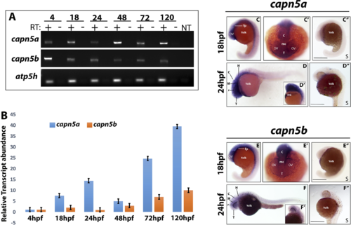
capn5a and capn5b are expressed in the developing brain of zebrafish. (A) RT-PCR for capn5a and capn5b expression during development (24, 48, 72, and 120 hpf). atp5h expression is shown as housekeeping gene control. RT, reverse transcriptase; NT, no template. (B) qPCR representation of fold-change in mRNA expression relative to 24 hpf. (C–C″) WISH of capn5a at 18 hpf. Strong capn5a expression was observed in the optic vesicles and brain. WISH with a control sense probe is shown. (C″, D–D″) WISH for capn5a at 24 hpf. Strong expression of capn5a was detected in the brain and hatching gland. Control sense probe is shown. (D″, E–E″) WISH for capn5b at 18 hpf. Expression was observed in parts of the developing brain but not the optic vesicles. Sense probe shown in (E″, F–F″) WISH for capn5b at 24 hpf. Modest expression of capn5b was observed in the brain. c, cerebellum; fp, floor plate; H, hindbrain; HG, hatching gland; OV, optic vesicle; mc, mesencephalon; r, retina; T, telencephalon. Scale bars: 50 μm.
|
Expression Data
| Genes: | |
|---|---|
| Fish: | |
| Anatomical Terms: | |
| Stage Range: | Sphere to Day 5 |
Expression Detail
Antibody Labeling
Phenotype Data
Phenotype Detail
Acknowledgments
This image is the copyrighted work of the attributed author or publisher, and
ZFIN has permission only to display this image to its users.
Additional permissions should be obtained from the applicable author or publisher of the image.
Full text @ Invest. Ophthalmol. Vis. Sci.