
Fig. 2
- ID
- ZDB-FIG-180828-11
- Publication
- Zhang et al., 2018 - Apoptosis of Endothelial Cells Contributes to Brain Vessel Pruning of Zebrafish During Development
- Other Figures
- All Figure Page
- Back to All Figure Page
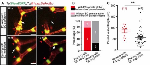
Characterization of EC apoptosis-accompanied pruned vessels. (A) In vivo time-lapse confocal images showing the relative spatial locations of EC nuclei in pruned and adjacent brain vessels. Top: for an EC apoptosis-accompanied pruned brain vessel, the nuclei of two neighboring vessels’ ECs (red arrowheads) located at the both ends of the pruned vessel, respectively (white arrow). Bottom: for an EC migration-accompanied pruned brain vessel, the nuclei of neighboring vessels’ ECs (red arrowheads) did not occupy the ends of the pruned vessel (white arrow). (B) Summary of data showing that EC apoptosis-accompanied pruning vessels have a higher probability that neighboring vessels’ EC nuclei occupy their both ends (7 out of 9, n = 7 larvae) than EC migration-accompanied pruning vessels (3 out of 11, n = 6 larvae). (C) Length of pruned vessels with EC apoptosis (n = 9 larvae) or EC migration (n = 13 larvae). The numbers on the bars (B) or in the brackets (C) represent the number of pruned vessels examined. Data are shown as mean ± SEM. **p < 0.01 (two-tailed unpaired Student’s t-test). Scale bar: 10 μm (A). |