
FIGURE
Fig. 8
- ID
- ZDB-FIG-180823-36
- Publication
- Bernstein et al., 2018 - The cellular bases of choroid fissure formation and closure
- Other Figures
- All Figure Page
- Back to All Figure Page
Fig. 8
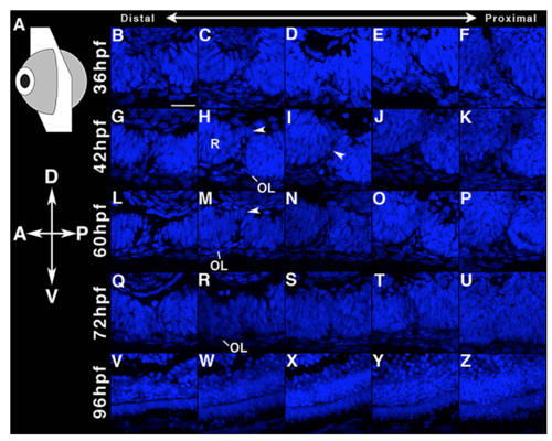
CF closure in the Zebrafish. Each image corresponds to the Laminin and DAPI co-labeled images shown in Fig. 7. The arrowheads in H and M mark the dorsal involution and apposition of the retinal folds. The arrowhead in I indicates the absence of retinal CF fusion despite BM continuity across the dorsal and ventral surfaces of the CF (See Fig. 7I). Scale bar: 20µm. |
Expression Data
Expression Detail
Antibody Labeling
Phenotype Data
Phenotype Detail
Acknowledgments
This image is the copyrighted work of the attributed author or publisher, and
ZFIN has permission only to display this image to its users.
Additional permissions should be obtained from the applicable author or publisher of the image.
Reprinted from Developmental Biology, 440, Bernstein, C.S., Anderson, M.T., Gohel, C., Slater, K., Gross, J.M., Agarwala, S., The cellular bases of choroid fissure formation and closure, 137-151, Copyright (2018) with permission from Elsevier. Full text @ Dev. Biol.