
- Title
-
The cellular bases of choroid fissure formation and closure
- Authors
- Bernstein, C.S., Anderson, M.T., Gohel, C., Slater, K., Gross, J.M., Agarwala, S.
- Source
- Full text @ Dev. Biol.

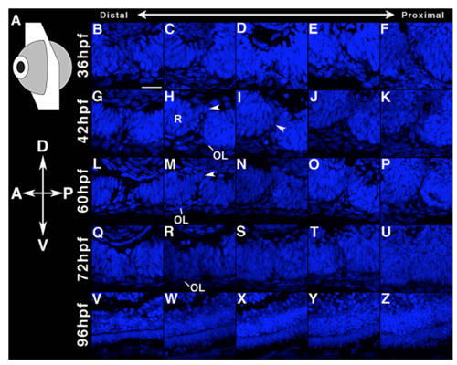
CF closure in the zebrafish. Lam and DAPI labeled tangential sections through the zebrafish CF at 36–96 hpf. Corresponding DAPI-only channels for each panel are provided in Fig. S8. Scale bar = 20 µm. A. Schematics for orientation. B–F. At 36 hpf, apposition occurs variably along the proximal-distal axis (arrowheads C, E). BM breakdown initiates at E (arrowhead). G, H. At 42 h, CF involution apposes the distal retinal folds dorsally and draws the OL to the center of the CF (arrowhead, H; magnified in inset, H,). BM breakdown in the retina occurs dorsal to the arrowhead. See also, Fig. S8G, H. I–K. The BM between medial CF folds has disappeared (I, J). Although the ILM and Bruch's membrane are continuous, the CF folds are not fused (magnified in inset, I; See also, Fig. S8I, J). BM breakdown (arrowhead, K) is also apparent proximally, ventral to the ON. L–P. At 60 hpf, the CF folds are separated distally (L, M). BM breakdown and ILM and Bruch's membrane continuity are established medially and proximally although tissue fusion has not yet occurred (N–P). See Fig. S8L–P. Q–U. At 72 hpf. CF closure is complete at all but distal locations (Q, R). The OL has retracted from the CF. However, the fusion of the retinal folds is complete only proximally (Fig. 7U) and not at other locations (Fig. 7T). See also, Fig. S8Q–U. V–Z. Fusion is complete at 96 hpf. |
CF closure in the Zebrafish. Each image corresponds to the Laminin and DAPI co-labeled images shown in Fig. 7. The arrowheads in H and M mark the dorsal involution and apposition of the retinal folds. The arrowhead in I indicates the absence of retinal CF fusion despite BM continuity across the dorsal and ventral surfaces of the CF (See Fig. 7I). Scale bar: 20µm. |
Reprinted from Developmental Biology, 440, Bernstein, C.S., Anderson, M.T., Gohel, C., Slater, K., Gross, J.M., Agarwala, S., The cellular bases of choroid fissure formation and closure, 137-151, Copyright (2018) with permission from Elsevier. Full text @ Dev. Biol.