
Fig. 3
- ID
- ZDB-FIG-180822-32
- Publication
- Schultz et al., 2018 - Epigenetic regulators Rbbp4 and Hdac1 are overexpressed in a zebrafish model of RB1 embryonal brain tumor, and are required for neural progenitor survival and proliferation
- Other Figures
- All Figure Page
- Back to All Figure Page
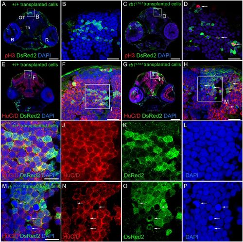
Cell-autonomous requirement for RB1 in blocking cell cycle entry in zebrafish neural precursors. Immunolabeling of 5 dpf host larval brain sections containing descendants of +/+; ubi:DsRed2 and rb1Δ7/Δ7; ubi:DsRed2 donor transplanted cells. n=3 individual transplant experiments for each donor genotype. (A-D) Phosphohistone H3- and DsRed2-labeled sections through the midbrain optic tectum (OT), thalamus (Th) and retinas (R). Wild-type (A,B) and rb1Δ7/Δ7 mutant (C,D) cells present in a wild-type host optic tectum (OT). A few phosphohistone H3-positive rb1Δ7/Δ7 mutant cells can be detected (D, arrows). (E-H) Neuronal marker HuC/D- and DsRed2-labeled sections. Wild-type (E,F) and rb1Δ7/Δ7 mutant (G,H) cells in a host optic tectum express HuC/D. A number of rb1Δ7/Δ7 mutant cells with highly condensed chromatin lack HuC/D labeling (H, arrows). (I-J) Boxed region in F, showing HuC/D- and DsRed2-positive wild-type cells with interphase chromatin. (M-P) Boxed region in H, showing that DsRed2-positive rb1Δ7/Δ7 mutant cells with interphase chromatin also express HuC/D. HuC/D is absent from rb1Δ7/Δ7 mutant cells with highly condensed chromatin (arrows). Scale bars: 100 µm (A,C,E,G); 20 µm (B,D,F,H); 10 µm (I,M). |