
Fig. 3-S1
- ID
- ZDB-FIG-180821-44
- Publication
- Fukui et al., 2018 - Hippo signaling determines the number of venous pole cells that originate from the anterior lateral plate mesoderm in zebrafish
- Other Figures
- All Figure Page
- Back to All Figure Page
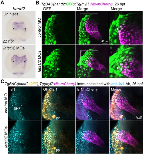
Depletion of Lats1/2 results in an increase in the number of hand2-promoter-active cells in the venous pole. (A) WISH analyses at 22 hpf of the control embryos (uninjected, n = 48) and embryos injected with lats1/2 MOs (n = 76) using an antisense probe for hand2. (B) 3D confocal stack images (at 26 hpf) of the TgBAC(hand2:GFP);Tg(myl7:Nls-mCherry) embryos injected with control MO (n = 12) and lats1/2 MOs (n = 16). GFP images (left), merged GFP and mCherry images (center), and enlarged images of the boxed regions in the center panels (right). Dorsal view, anterior to the top. Images are a set of representative images from eight independent experiments. (C) Confocal 3D-stack images (at 26 hpf) of the TgBAC(hand2:GFP);Tg(myl7:Nls-mCherry) embryos injected with the MOs indicated on the left and immunostained with the anti-Isl1 Ab (control MO, n = 15; lats1/2 MOs, n = 21). Square brackets denote the SHF cells that are Isl1-positive. The first, second, third and fourth images are Isl1 immunostaining, the merged GFP and Isl1 immunostaining image, the merged Isl1 immunostaining and mCherry image, and the merged image for all the three labels (GFP, mCherry, and Isl1 immunostaining), respectively. pp indicates the pharyngeal pouch, which expresses hand2-promoter-activated GFP signals. Dorsal view, anterior to the top. The images are a set of representative images for three independent experiments. |