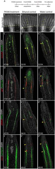
Inhibition of calcineurin activity suppresses joint formation and joint-related gene expression. (A) Fish were treated with FK506 for 2 days prior to amputation. At 0 dpa/2 dot, fins were amputated and treatments continued for 4 days. (B) At 4 dpa/6 dot, no joints form in the regenerates of FK506-treated fish. (B′,B″) Ethanol (B′) and water (B″) controls form joints normally in the regenerate (yellow arrowheads). Dashed yellow lines indicate amputation plane. (C-F″) Double FISH on longitudinal cryosections of 4 dpa regenerates. (C) In FK506-treated fins, runx2a and runx2b are expressed, but joint cell clusters are absent (no ‘bump’ in the expression domain). (D-F) FK506 treatment inhibits hoxa13a (D), pthlha (E) and evx1 (F) expression. sp7 (D), ihha (E) and col10a1 (F) domains of expression are uninterrupted in committed osteoblasts owing to the absence of joints. However, sp7 is still absent in the distal pre-osteoblasts (D, pink bracket). (C′-F″) Expression of joint (yellow arrowheads) and osteoblast markers are unaffected in ethanol (C′-F′) and water (C″-F″) controls. Numbers in each panel represent the number of sections with the expression pattern over the total number of sections analyzed. Scale bars: 500 μm (in B for B-B″); 50 μm (in C for C-F″).
|
