
FIGURE
Fig. 3
- ID
- ZDB-FIG-180806-4
- Publication
- Desvignes et al., 2018 - Evolution of caudal fin ray development and caudal fin hypural diastema complex in spotted gar, teleosts, and other neopterygian fishes
- Other Figures
- All Figure Page
- Back to All Figure Page
Fig. 3
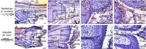
Orcein stains for elastin in plates of connective tissue in spotted gar and zebrafish. Sagittal sections of caudal fin regions of (A) a 24 mm TL spotted gar larva (∼22 dpf) and (B) a 13 mm TL zebrafish (21 dpf) stained with Orcein (red‐rusty color) and counter stained with Gills hematoxylin (blue‐purple). Scale bar = 100 μm. White elongated triangles indicate hypural 1 and black arrows point at the hypural diastema. hd, hypural diastema; hyp, hypural; nc, notochord; oc, opisthural cartilage; pcta, anterior plate of connective tissue; pctp, posterior plate of connective tissue; phy, parhypural; rbc, red blood cells; vas, vasculature.
|
Expression Data
Expression Detail
Antibody Labeling
Phenotype Data
Phenotype Detail
Acknowledgments
This image is the copyrighted work of the attributed author or publisher, and
ZFIN has permission only to display this image to its users.
Additional permissions should be obtained from the applicable author or publisher of the image.
Full text @ Dev. Dyn.