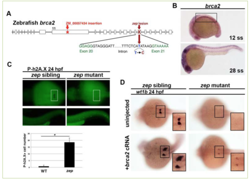
brca2 is necessary and sufficient to promote podocyte development in the embryonic zebrafish kidney. A) Whole genome sequencing (WGS) revealed that zep mutants contain a T to C transversion in the brca2 gene that we found resulted in aberrant splicing. This schematic map also indicates the location of the lesion in brca2ZM_00057434 insertional mutants used in our study. B) WISH on wild-type fish at a variety of stages showed that brca2 transcripts (purple stain) were expressed ubiquitously throughout the organism, including the intermediate mesoderm (IM) which gives rise to kidney (boxed area). C) zep siblings and mutants at the 24 hpf time point were subjected to WISH with wt1b to demarcate podocytes (boxed area), then immunofluorescence to detect phosphorylated h2A.X (P-h2A.X). There was a significant increase in cells positive for P-h2A.X in zep mutants, which indicates an accumulation of double stranded DNA breaks, and suggested that zep mutants have dysfunctional brca2 protein (* = p<0.05). D) Both zep siblings and zep mutants were injected with brca2 capped RNA (cRNA) and then subjected to WISH with wt1b. Interestingly, some injected wild-type and heterozygous sibling embryos had drastically increased areas of podocytes (frequency of 13.5%) compared to uninjected controls. Podocyte development was partially rescued in zep mutants (frequency of 55%) following brca2 overexpression.
|
