
FIGURE
Fig. 7
- ID
- ZDB-FIG-180712-58
- Publication
- Chawla et al., 2018 - Retinoic Acid Maintains Function of Neural Crest-Derived Ocular and Craniofacial Structures in Adult Zebrafish
- Other Figures
- All Figure Page
- Back to All Figure Page
Fig. 7
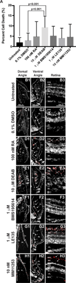
RA regulates cell survival in the anterior segment and retina. TUNEL assay showed that 2-day treatment with 10 μM DEAB (9.3% ± 5.4%) significantly increased the percentage of apoptotic cells (A) within the adult zebrafish eye as compared to untreated (2.0% ± 2.4%, P < 0.001) and 0.1% DMSO control–treated fish (3.0% ± 3.5%, P < 0.001). Two-day treatment with 100 nM RA (4.8% ± 7.3%), 1 μM BMS195614 (4.1% ± 4.8%), 1 μM LE135 (5.2% ± 4.6%), or 10 nM MM11253 (4.6% ± 6.2%) did not significantly change the percentage of apoptotic cells in the zebrafish eye. Despite no significant change in percentage of apoptotic cells in the eye, apoptosis was localized in RA-treated fish to the IS, canalicular network (arrows), and aqueous plexus (arrowheads) in the ventral iridocorneal angle (D2) as compared to untreated (B2) and DMSO control–treated (C2) fish. RA also showed increased apoptosis within the retinal pigment epithelium (arrows, [D3 compared to B3 and C3]), but did not affect cell survival in the dorsal iridocorneal angle (D1 compared to B1 and C1). Treatment with DEAB (10 μM) for 2 days caused diffuse apoptosis in the dorsal (E1) and ventral (E2) iridocorneal angles and in the retinal pigment epithelium and photoreceptors of the retina (arrows, [E3]). Treatment with the RARα-specific antagonist BMS195614 (1 μM) for 2 days did not affect cell survival in the dorsal angle (F1), ventral angle (F2), or the retina (F3). Inhibition of RARβ with LE135 (1 μM) for 2 days did not increase apoptosis in the angles (G1, G2) but did increase apoptosis in the retina (arrows, [G3]). The RARγ antagonist MM11253 (10 nM) caused localized apoptosis in the ventral angle (H2) and retina (H3), but not the dorsal angle (H1). AL, annular ligament; Co, cornea; GCL, ganglion cell layer; INL, inner nuclear layer; ONL, outer nuclear layer.
|
Expression Data
Expression Detail
Antibody Labeling
Phenotype Data
Phenotype Detail
Acknowledgments
This image is the copyrighted work of the attributed author or publisher, and
ZFIN has permission only to display this image to its users.
Additional permissions should be obtained from the applicable author or publisher of the image.
Full text @ Invest. Ophthalmol. Vis. Sci.