
FIGURE
Fig. 3
- ID
- ZDB-FIG-180712-55
- Publication
- Chawla et al., 2018 - Retinoic Acid Maintains Function of Neural Crest-Derived Ocular and Craniofacial Structures in Adult Zebrafish
- Other Figures
- All Figure Page
- Back to All Figure Page
Fig. 3
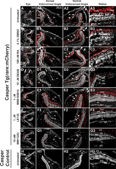
RA activity is in the retina and iridocorneal angles in the adult eye. In Casper Tg(rare:mCherry) adult zebrafish, RA activity was predominantly in the PRs and RPE in the adult eye of untreated (A, A3) and 0.1% DMSO control–treated (B, B3) fish. RA was also found in the OPL and IPL (A3, B3) in the retina. In the dorsal iridocorneal angle of untreated and control-treated eyes (A1, B1), RA activity was in the IS. In the ventral iridocorneal angle of untreated and control-treated eyes (A2, B2), RA was found in IS, the canalicular network that was between the iris and annular ligament (arrows), and the angular aqueous plexus (arrowheads). There was no detectable RA activity in the cornea (Co) in this reporter line. Treatment with 100 nM RA for 5 days (C) increased RA activity in the IS in the dorsal (C1) and ventral (C2) angles and in the ventral canalicular network (arrows, [C2]), but not in the retina (C3). Treatment with 10 μM DEAB for 5 days (D) decreased RA activity throughout the eye including in the dorsal (D1) and ventral (D2) angles and in the retina (D3). Treatment with the RARα-specific antagonist BMS195614 (1 μM) for 5 days did not alter RA activity within the eye (E, E1, E2, E3). Treatment with the RARβ antagonist LE135 (1 μM, F) for 5 days showed mildly decreased RA activity in the retina in the PRs, OPL, and IPL (F3), but no change in the dorsal (F1) or ventral (F2) angles. Inhibition of RARγ with the specific antagonist MM11253 (10 nM) decreased RA throughout the eye (G), including in the angles (G1, G2) and retina (G3). Control Casper fish, which lack the transgene, showed minimal to no fluorescence in the eye (H, H1, H2, H3). AL, annular ligament; GCL, ganglion cell layer; INL, inner nuclear layer; ONL outer nuclear layer.
|
Expression Data
Expression Detail
Antibody Labeling
Phenotype Data
Phenotype Detail
Acknowledgments
This image is the copyrighted work of the attributed author or publisher, and
ZFIN has permission only to display this image to its users.
Additional permissions should be obtained from the applicable author or publisher of the image.
Full text @ Invest. Ophthalmol. Vis. Sci.