
FIGURE
Fig. 3-S1
- ID
- ZDB-FIG-180712-5
- Publication
- Viader-Llargués et al., 2018 - Live cell-lineage tracing and machine learning reveal patterns of organ regeneration
- Other Figures
- All Figure Page
- Back to All Figure Page
Fig. 3-S1
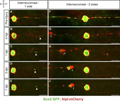
Interneuromast cells do not regenerate. (A–E) The ablation of interneuromast cells adjacent to one flank of a neuromast resulted in the stretching of the last undamaged interneuromast cell (arrowhead) but does not trigger interneuromast-cell proliferation or the reformation of interneuromast-cell strings (n = 14). (F–J) Likewise, the complete ablation of interneuromast cells in both flanks from one neuromast to the next, generates a corresponding gap of interneuromast cells that did not change over 7 days (n = 8). Scale bars: 50 µm. |
Expression Data
Expression Detail
Antibody Labeling
Phenotype Data
Phenotype Detail
Acknowledgments
This image is the copyrighted work of the attributed author or publisher, and
ZFIN has permission only to display this image to its users.
Additional permissions should be obtained from the applicable author or publisher of the image.
Full text @ Elife