
Fig. S3
- ID
- ZDB-FIG-180702-24
- Publication
- Bergboer et al., 2018 - Assaying sensory ciliopathies using calcium biosensor expression in zebrafish ciliated olfactory neurons
- Other Figures
- All Figure Page
- Back to All Figure Page
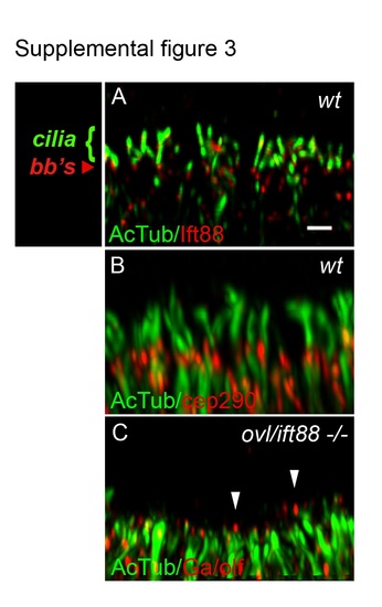
Olfactory sensory cilia deficit in oval/ift88 -/- mutants. (A) Olfactory sensory cilia in the center of a wild type olfactory placode stained with anti-Ift88 (red) and anti-acetylated tubulin (green; cilia) and imaged in confocal Z-stacks. Ift88 immunoreactivity was strongest in basal bodies (bb's). Scale bar in (A) equals 1 µm. (B) Wild type olfactory placode stained with anti-acetylated tubulin (green; cilia and neuronal cell body processes) and anti-cep290 (red; basal bodies). (C) oval mutant olfactory placode stained with anti-G /olf (red) and anti-acetylated tubulin (green) shows cilia loss with some short, residual G /olf-positive axonemes (arrowheads). All panels are set at equivalent scale and represent a 3µm thick maximum intensity projection of the center of the olfactory placode. |