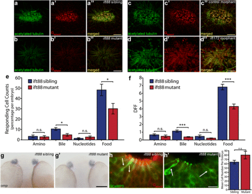
Cilia defects have functional effects in OSNs in Ift-deficient zebrafish at 2.5 dpf. a Anti-acetylated tubulin (green) stains all cilia axonemes in wildtype embryos as well as neuronal processes projecting away from the olfactory placode. a’ Anti-Ga/olf (red) is a marker for OSN cilia. a” Merged image of a and a’. b ift88 mutant homozygotes show loss of acetylated tubulin positive axonemes and b’ loss of anti-Ga/olf staining. b” Merged image of b and b’. c Control morpholino injected olfactory placode stained with anti-acetylated tubulin and c’ anti-Ga/olf. c” Merged image of c and c’. d ift172 morphant olfactory placodes show loss of anti-acetylated tubulin staining and d’ loss of anti-Ga/olf staining. d” Merged image of d and d’. Remaining anti-acetylated tubulin staining is restricted to neuronal processes [67] (b, d). For quantification see Table 1. e Significant change in percentage of odour responding OSNs per embryo after addition of bile acids and food odour in Tg(elavl3:GCaMP5) ift88 mutant OSNs compared to sibling OSNs (N = 637 OSNs, 10 fish for sibling and N = 720 OSNs, 9 fish for mutant). f Significantly reduced response amplitude to bile acids and food odour in Tg(elavl3:GCaMP5) ift88 mutant responding OSNs compared to sibling responding OSNs. g Whole mount in situ hybridization using an omp probe demonstrates clear omp expression in the OE in both wild type siblings g and g’ ift88 mutants and siblings at 2 dpf. (For other time points see Additional file 1: Fig. S6A). h Ciliated OSNs stained with anti-GFP (green) and anti-Ga/olf (red) present in both h sibling and h’ ift88 mutant, based on cell shape (arrows) in Tg(elavl3:GCaMP5) fish. i No difference in number of GCaMP5 positive OSNs per fish (P = 0.10). Bars represent mean and SEM (*P < 0.05, **P < 0.01, ***P < 0.001 Mann–Whitney U test for e and f, student’s t test for i. Scale bar is 10 μm, except panel g bar is 100 μm
|
