
Fig. S5
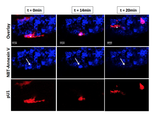
Microglial engulfment of apoptotic neurons at 72 hpf. Representative static images from live imaging of a microglial cell engulfing an apoptotic neuron in the spinal cord at 72 hpf following BPA exposure in a double transgenic Tg:pU1:Gal4-UAS-RFP/NBT:DLexPR-secA5-TagBFP embryo, which have pU1+ microglia cells labeled in red and apoptotic neurons labeled in blue. Asterisks denotes pU1+ cell (in red) in the process of engulfing a neuron undergoing apoptosis (in blue) in the spinal cord and its subsequent migration away from this site. White arrow denotes an apoptotic neuron being removed from the spinal cord over a time course of 20 minutes. Note that specific microglia were not followed from 48 hpf to 72 hpf, and cells imaged in this figure illustrate the general microglial response in the spinal cord at 72 hpf. Images were collected from N = 1 biological replicate. |