
Fig. 3
- ID
- ZDB-FIG-180626-21
- Publication
- Takei et al., 2018 - High-Sensitivity and High-Resolution In Situ Hybridization of Coding and Long Non-coding RNAs in Vertebrate Ovaries and Testes
- Other Figures
- All Figure Page
- Back to All Figure Page
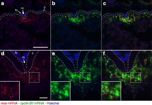
Double fluorescence in situ hybridization of mos (red) and cyclin B1 (green) mRNAs in zebrafish ovaries. DNA is shown in blue. a-c A zebrafish ovary section showing localization of mos (a) and cyclin B1 (b) mRNAs in a fully grown oocyte. A merged image is shown in (c). The mos and cyclin B1 mRNAs were localized at the animal polar cytoplasm beneath the micropyle (m). fc, follicle cells; c, chorion. d-f High resolution imaging of the oocyte shown in a-c. The insets are enlarged views of the boxed regions. The mos and cyclin B1 mRNAs were distributed in the animal polar cytoplasm of oocyte as different granules. Bars: 50 μm in a-c, 10 μm in d-f |
| Genes: | |
|---|---|
| Fish: | |
| Anatomical Term: | |
| Stage: | Adult |