
Fig. 2
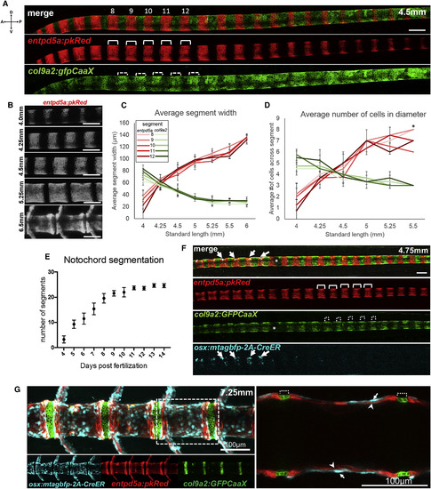
The Notochord Sheath Segments into Alternating Domains Prior to Spine Morphogenesis (A) Live confocal images showing alternating entpd5a:pkRED (solid brackets) and col9a2:GFPCaaX segments (dotted brackets) in the notochord sheath of a 4.5 mm SL fish. Numbers represent the entpd5a segments used for quantifications. (B) At 4.0 mm SL, entpd5a:pkRED expression produces segments that expand over time and completely mirror centra. (C) entpd5a+ segment length (red tones), monitored in segments 8–12, increases as col9a2+ domain (green tones) length decreases in three individual fish. (D) The number of cells within entpd5a+ segments increase via the transition of adjacent col9a2+ cells were measured in three individual fish. ∗Cells could not be counted accurately because of crowding. (E) Monitoring the appearance of entpd5a+ segments over time indicates new segments form sequentially every 8 hr. Between 15 and 32 fish were quantified for each time point. Means and SDs are displayed for each time point. (F) Confocal image of a 4.75 mm SL fish showing that segregation of alternating entpd5a+ (solid brackets) and col9a2+ (dotted brackets) segments in the notochord sheath precedes osteoblast recruitment (cyan; marked by osx), which are observed only in the oldest, most anterior segments (denoted by arrows). (G) Confocal image of a 7.25 mm SL fish showing that entpd5a:pkRED expression (arrowheads) underlies osteoblasts, marked by Tg(osx:mTagBFP-2A-CreER) expression, in the spine centra (arrows), while col9a2:GFPCaaX expression is confined to the IVD (dotted brackets). Right: a zoomed-in z-slice of dotted box in left image. Developmental stages are based on standard length. Scale bars are 100 μm. Asterisk denotes interference in optical path. Images in (A), (B), and (F) are digitally stitched. See also Figure S1. |