
Fig. 7
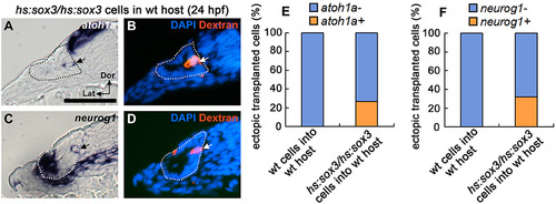
Effects of early misexpression ofsox3in genetic mosaics. (A–D) Expression of atoh1a (A) and neurog1 (C) at 24 hpf in cross sections of wild-type hosts into which fluorescent dextran-labeled hs:sox3/hs:sox3 transgenic cells were transplanted. Mosaic embryos were heat shocked at 12.5 hpf, 38 °C for 30 min. (B, D) Fluorescent image of dextran and DAPI staining on the same sample shown in A and C respectively. Sections pass through the middle of the otic vesicle, just posterior to the utricular macula. Arrows indicate transgenic cells that ectopically express atoh1a or neurog1. Otic vesicle borders are outlined. (E, F) Quantification of the percentage of transgenic cells located outside endogenous sensory or neural domains that ectopically express atoh1a (E) or neurog1 (F). |
Reprinted from Developmental Biology, 435(1), Gou, Y., Vemaraju, S., Sweet, E.M., Kwon, H.J., Riley, B.B., sox2 and sox3 play unique roles in development of hair cells and neurons in the zebrafish inner ear, 73-83, Copyright (2018) with permission from Elsevier. Full text @ Dev. Biol.