
Fig. S3
- ID
- ZDB-FIG-180611-72
- Publication
- Freudenblum et al., 2018 - In vivo imaging of emerging endocrine cells reveals a requirement for PI3K-regulated motility in pancreatic islet morphogenesis.
- Other Figures
- All Figure Page
- Back to All Figure Page
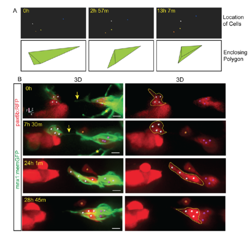
Protrusion formation coincides with endocrine cell movements and clustering. Related to Figure 2. (A, top) Tracked cells (colored spheres) shown alone, from the time series shown in Fig 2B. (A, bottom) Enclosing polygon as calculated using Matlab. Polygon volume reflects the spread of cells in three-dimensional space, and thus provides an index of cell clustering. (B) Image series of pancreatic endocrine cells in pax6b:dsRed;mnx1:memGFP transgenics following Notch inhibitor treatment from 4 dpf to 5 dpf, and imaged at 6 dpf. 3D representations with cells tracked (colored spheres) using Imaris. Movement of cells follows the appearance of a fine intercellular tether (arrow). Scale bars = 10μm. *Indicates neurite projections from mnx1:memGFP transgene expressed in overlying neurons. |