
Fig. 1
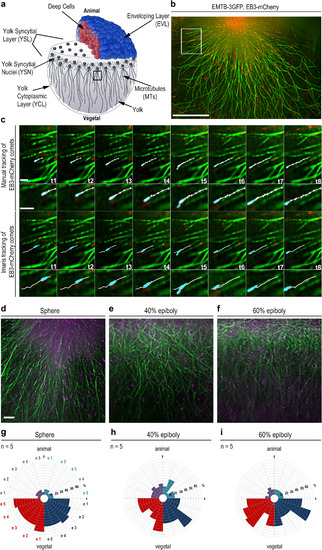
Visualization of yolk cell microtubule dynamics. (a) Wild-type embryo at sphere stage (4 hpf). Embryo images in all figures are shown in lateral views, animal at top. Black square: YCL region beneath YSL, in which microtubule growth dynamics was analyzed. (b) Live sphere stage wild-type embryo expressing EMTB-3GFP (green) and EB3-mCherry (red) imaged by TIRF microscopy in the region marked in (a). Scale bar 20 µm. (c) Time series of microtubules of the region marked in (b). EB3-mCherry (cyan) tracking over 8 time points (1.7 s per frame). Top rows manual tracking (white, one cross per frame), bottom rows Imaris® tracking. Higher magnifications of the tracks in the small images. Both tracking methods yielded similar track lengths (manual 4.77 µm +/− 0.18 stdev; Imaris 4.71 µm +/− 0.05). Scale bars 2 µm. (d-f) YCL regions of EMTB-3GFP (green) and EB3-mCherry (magenta) labeled live wild-type embryos at indicated developmental stages. Scale bar 5 µm. (g-i) Directional distribution of EB3-mCherry tracks binned into 90 degree color coded quadrants. Each embryo (e1-e5) shown as one segment per quadrant. A significantly higher number of tracks have vegetal direction at all stages (p<0.005; Mann-Whitney test). |
Reprinted from Developmental Biology, 434(2), Eckerle, S., Ringler, M., Lecaudey, V., Nitschke, R., Driever, W., Progesterone modulates microtubule dynamics and epiboly progression during zebrafish gastrulation, 249-266, Copyright (2017) with permission from Elsevier. Full text @ Dev. Biol.