
Fig. 3
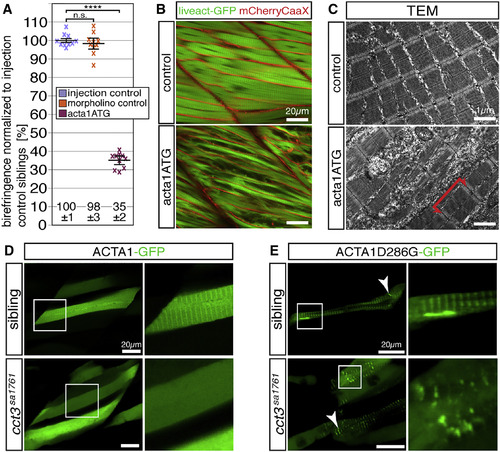
Actin Folding Is Impaired in cct3sa1761 (A) Injection of 3-dpf-old WT larvae with 100 μM acta1ATG morpholino targeting acta1a and acta1b significantly reduced their birefringence in comparison with larvae injected with injection solution (injection control) or 100 μM standard control morpholino (morpholino control). Data are mean ± SEM; ∗∗∗∗p < 0.0001 by Student’s t test; n = 10. (B) The reduced amount of myofibrils after acta1a and acta1b knockdown was also documented in the transgenic background of Tg(acta1:mCherryCaaX) and Tg(acta1:lifeact-GFP) (n = 4 per genotype). (C) TEM revealed that knockdown of acta1a and acta1b reduced the amount of myofibrils but did not affect sarcomere organization (n = 3 per genotype). (D) After incorporation of ACTA1-GFP, the myofibrils of siblings presented a striated fluorescence pattern, whereas the fluorescence of the cct3sa1761 myofibrils appeared uniform (4 larvae per genotype, each with 5 myofibers analyzed). (E) Mutant ACTA1D286G-GFP formed rod-shaped structures (arrowhead) in siblings, whereas, in cct3sa1761 homozygotes, exclusively amorphic aggregates of various sizes (arrowhead) were observed (4 larvae per genotype, each with 5 myofibers analyzed). See also Figure S6 and Table S1. |
| Genes: | |
|---|---|
| Fish: | |
| Knockdown Reagent: | |
| Anatomical Terms: | |
| Stage: | Protruding-mouth |
| Fish: | |
|---|---|
| Knockdown Reagent: | |
| Observed In: | |
| Stage: | Protruding-mouth |