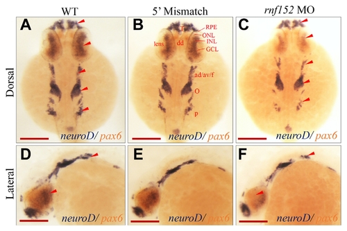
Two color WISH analysis with neuroD- and pax6–specific probes detected changes in their transcripts in the retinal layers of embryos at 27 hpf. (A, D) WT, (B, E) 5′ mismatch MO control, and (C, F) rnf152 MO. neuroD transcripts (dark blue) were abundant in the INL and ONL of eyes, telencephalon, dorsal diencephalon, octaval/statoacustic ganglia and posterior lateral line ganglia of WT as well as the 5′ mismatch control, but remarkably reduced in rnf152 MO. Levels of pax6 transcripts (tomato color) in the telencephalon, dorsal diencephalon, hindbrain, anterior spinal cord, and eyes of the WT were similar those of rnf152 MO at 27 hpf. Red arrowheads indicates the areas of significant differences in the level of neuroD transcripts between WT and rnf152 MO. Abbreviations: ad/av/f- Anterodorsal/anteroventral lateral line/facial placodes/ganglia, dd- Dorsal diencephalon, GCL- Ganglion cell layer, INL- Inner nuclear layer, o- Octaval/statoacustic ganglia. ONL- outer nuclear layer, p- Posterior lateral line ganglia, RPE- retinal pigment epithelium, T- Telencephalon (n = 3). Scale bars (A–F): 50 μm.
|
