
Fig. 1-S1
- ID
- ZDB-FIG-180504-13
- Publication
- Weber et al., 2017 - Cell-accurate optical mapping across the entire developing heart
- Other Figures
- All Figure Page
- Back to All Figure Page
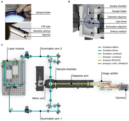
High-speed light sheet microscopy for in vivo 3D optical mapping. (A) A zebrafish embryo is mounted in agarose inside a fluorinated ethylene propylene (FEP) tube. (B) Section view of the sample holder with mounted zebrafish embryo placed inside the medium-filled sample chamber. The embryo is placed in the field of view of the detection objective and illuminated with a static light sheet from one of two sides. (C) Top view of the high-speed light sheet microscope for in vivo cardiac imaging. The laser module combines a 488 and a 561 nm laser line and sends the beam into the two illumination arms. Both arms generate identical light sheets from two opposite sides. The motor unit positions the sample holder with the mounted zebrafish embryo at the intersection of illumination and detection path. Fluorescence emission is split and recorded with an sCMOS camera running at up to 400 Hz. |