
Fig. S2
- ID
- ZDB-FIG-180502-22
- Publication
- Rabani et al., 2017 - A Massively Parallel Reporter Assay of 3' UTR Sequences Identifies In Vivo Rules for mRNA Degradation
- Other Figures
- All Figure Page
- Back to All Figure Page
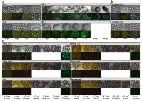
Development of zebrafish embryos after injection of mRNA reporters. Related to Figure 1. Images of zebrafish embryos or oocytes at different developmental stages (columns; developmental times as indicated on bottom) after they were injected with mRNA reporters at the 1-cell stage. Top panels show an image of the developmental stage, and lower panels show GFP florescence (green) that arise from translation of the mRNA reporters in those embryos. (a) Embryos injected with A+ (top) or A- (bottom) reporters. (b) Oocytes injected with A+ (top) or A- (bottom) reporters. (c) Embryos injected with 80pg, 20pg or 0pg of A+ (left) or A- (right) reporters and uninjected controls. |