
FIGURE
Fig. S2
- ID
- ZDB-FIG-180420-46
- Publication
- Richter et al., 2017 - Small molecule screen in embryonic zebrafish using modular variations to target segmentation
- Other Figures
- All Figure Page
- Back to All Figure Page
Fig. S2
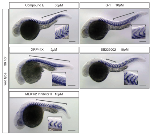
1st class segmentation phenotypes Identified hit phenotypes with direct segmentation defects according the classification used in Table 1, visible at 36 hpf after xirp2a in situ hybridization. All treatments are as labelled in the panel above the image. Anterior to the right, posterior to the left. Insets show zoom-in of the segment pattern and defects. Brackets indicate the area of the axis where segment boundary defects occurred. |
Expression Data
Expression Detail
Antibody Labeling
Phenotype Data
Phenotype Detail
Acknowledgments
This image is the copyrighted work of the attributed author or publisher, and
ZFIN has permission only to display this image to its users.
Additional permissions should be obtained from the applicable author or publisher of the image.
Full text @ Nat. Commun.