
Fig. 2
- ID
- ZDB-FIG-180417-20
- Publication
- Rogers et al., 2017 - Nodal patterning without Lefty inhibitory feedback is functional but fragile
- Other Figures
- All Figure Page
- Back to All Figure Page
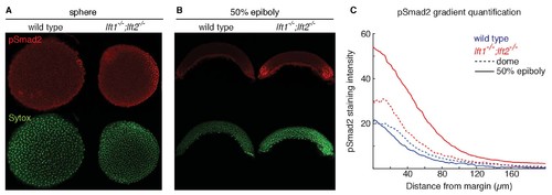
Lefty loss causes expanded Nodal signaling gradients. α-pSmad2 staining (red) at sphere stage (A, animal views) and 50% epiboly (B, lateral views) in representative wild type and lft1-/-;lft2-/- mutant embryos (nuclei labeled with Sytox nuclear stain (green)). Note stronger pSmad2 staining in lft1-/-;lft2-/- mutant embryos compared to wild type. (C) Quantification of pSmad2 gradients in wild type (blue) and lft1-/-;lft2-/- (red) mutant embryos at dome stage (dashed) and 50% epiboly (solid) shows an increase in the amplitude and range of the signaling gradient in lft1-/-;lft2-/- mutants. See Materials and methods and Figure 2—figure supplement 1 for quantification details. |
| Antibody: | |
|---|---|
| Fish: | |
| Anatomical Term: | |
| Stage Range: | Sphere to 50%-epiboly |
| Fish: | |
|---|---|
| Observed In: | |
| Stage Range: | Sphere to 50%-epiboly |