
Fig. 2
- ID
- ZDB-FIG-180417-1
- Publication
- Marsden et al., 2017 - The Nkd EF-Hand Domain Modulates Divergent Wnt Signaling Outputs in Zebrafish
- Other Figures
- All Figure Page
- Back to All Figure Page
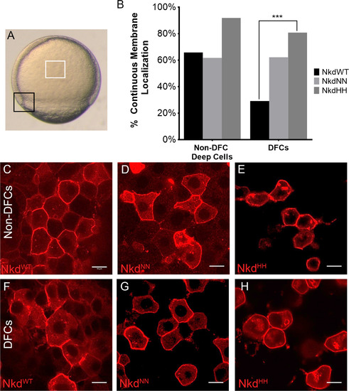
Nkd localization changes in calcium fluxing and quiescent cells. (A) Calcium-fluxing dorsal forerunner cells (DFCs) (black box) and calcium-quiescent non-DFC deep cells (white box) used for localization studies. (B) Analysis of continuous versus discontinuous membrane localization of all Nkd forms in the non-DFC deep cells and the DFCs. NkdWT shows a different localization pattern than NkdNN and NkdHH, which remain mostly membrane-bound. (C and F) NkdWT, NkdNN (D and G), and NkdHH (E and H) localization in the non-DFCs (C-E) and the DFCs (F-H). (F) Quantification of membrane localization. NkdWT shows a significantly different change in localization. ****= P-value<1×10⁻⁵, Fisher's exact test, two tail. NkdWT non-DFC n=120, NkdNN non-DFC n=94, NkdHH non-DFCs n=62, NkdWT DFC n=106, NkdNN DFC n=106, NkdHH n=52, n= number of cells. Scale bar 10 µm. Taken at 63x at 2x zoom. See also Fig. S3. |
Reprinted from Developmental Biology, 434(1), Marsden, A.N., Derry, S.W., Schneider, I., Scott, C.A., Westfall, T.A., Brastrom, L.K., Shea, M., Dawson, D.V., Slusarski, D.C., The Nkd EF-Hand Domain Modulates Divergent Wnt Signaling Outputs in Zebrafish, 63-73, Copyright (2017) with permission from Elsevier. Full text @ Dev. Biol.